
Introduction
The Value of a horse depends upon its ability to perform work. Four sound feet are indispensable to this end. An old adage among horse owners is "no foot-no horse".
In the wild, horses were practically free of serious foot problems. With thier domestication foot problems began to appear. Examining the changes in their environment, one can readily understand the causes. Horse were brought from relatively soft pastures to hard roads; from self-regulated exercise to enforced work; from healthy pastures to unsanitary barns where they were often forced to stand in feces, urine, and mud; from light self-limiting maintenance rations to heavy artificial diets, which are necessary for hard work.
Sound horses will frequently develop foot problems under an artificial environment and the misguided care of man. The possiblilites are even greater for horses haveing conformation defects. Therefore, it is important for the horse farm employee to carefully consider the priciples involved in properly caring for the hoofs and the shoeing of horses.
Reasons for Shoeing
Shoes protect the hoofs from excessive wear, provides better traction, help correct defects of stance or gait, help cure diseased or defected hoof(contracted heels, thrush, divided tendons), and may be used to provide relief from the pain of injured parts(hoof wall cracks, bruised soles, and tendonitis).
Although shoeing is a necessary evil for working horses, there are some misconceptions about the benefits of shoeing horses. The aplication of shoes does not make walking easier; the added weight of the shoes does not improve agility; and shoeing increases shock and road concussion. Nail holes made in attaching the shoe weaken the hoof wall, may cause separation, and may provide entry for infection.
Allowing a horse to wear the same shoes too long without trimming the hoofs and adjusting invites trouble. Since the walls of the hoof grow perpendicular to the coronary band, the base of support actually grows out from under the horse if the shoes are left on too long. This puts excessive strain on the flexor tendons. Also, shoes worn too long may become thin and loose and bend. They also may shift and cause shoe nail punctures or corns.
Structure and Action of the Foot
A knowledge of the anatomy of the feet and legs of a horse is essential for the proper care and shoeing.
Internal and External Parts of the Leg
Cannon Bone (Large Metacarpal Bone)
The cannon bone extends from the knee or hock to the
fetlock, is cylindrical in shape and stands upright.
The upper end is flat to form a large working surface
for the bones of the knee or hock. Its lower end has
an articular surface with three ridges, sepearated by
grooves. The middle prominence is the highest and
extends furthest forward. This articular surface is in
contract with the long Pastern Bones and Sesamoid
Bones. At teh sides of the lower end of the cannon
bone are two rough surfaces for the attachments of
ligaments.
Splint Bones (Internal & External Small Metacarpal Bones)
The splint bones are incompletely developed long bones
and are located one on each side, on the upper rear
portion of the cannon bones. The upper ends of the
splints are largest to form a more generous table for
the bones of the joint above, either the knee or the
hock to rest upon. The splints taper and terminate at
the lower portion of the cannon bone, shaped somewhat
like icycles. Fusion of the middle part of the shaft
with the cannon bone is common. The splints are
arranged on the cannon bone so that a furrow us formed
for containing ligaments and tendons.
Long Pastern Bone (First Phalanx)
The long pastern bone is about one-third the length of
the cannon bone. Its upper surface has three grooves
to accept the lower end of the cannon bone. The
configuration of the cannon-long pastern joint creates
a "perfect joint" that allows for no lateral movement.
The exterior of the bone is smooth, except at either
side of the upper portion, and an area on the
underside of the bone. These roughened surfaces create
an area for better attachment of ligaments. The lower
end of the long pastern has only one depression in its
articular surface.
Sesamoid Bones
The two sesamoid bones are shaped like pyramids. They
are located at the upper rear portion of the
articulatory surface of the long pastern bone. The
sesamoids are attached to the long pastern bone by
ligamentous tissue, and create a larger surface for
the rotation of the cannon-pastern joint.
Short Pastern Bone (Second Phalanx)
The short pastern or coronary bone is cube shaped, and
is about one half the size of the long pastern bone.
Its upper end is concave and shows two depressions to
receive the long pastern bone. The upper end is rather
large and has attachments for tendons. The lower end
has one depression in its convex articular
surface.
Coffin Bone (Third Phalanx)
The coffin bone is so named, perhaps, because it is
encased within the hoof. Its shape resembles the hoof.
The coffin bone is extremely light in comparison with
the other bones, and is perforated with many holes.
These wholes contain blood vessels and nerves that
infest the foot. The lightness of the coffin bone
allows the animal to use less power in moving the
legs.
The wall surfaces encompasses the front and sides
of the coffin bone. It is roughened to aid in
attachment of the sensitive lamina which covers it. At
the top of the wall surface in the center is a
projection, called the Extensor Process to which the
extensor tendon is attached. There are extensions to
the rear portion of the coffin bone called the wings
or Basilar processes.
The lower, sole surface of the coffin bone is
concave and smooth except at the rear portion. This
rear portion is roughened for the attachment of the
deep flexor tendon. This is called the tendinous
surface, or semi-lunar crest.
The articulatory surface has a prominence in the
middle of its concave surface and a depression on each
side of this ridge. It is formed to accept the lower
end of the coronary bone.
Navicular Bone
The navicular bone is shaped somewhat like a boat and
is situated between the wings of the coffin bone. This
bone is triangular in cross-section and is attached to
the coffin bone by ligamentous tissue. Its upper
surface corresponds to the articulatory surface of the
coffin bone and creates a larger rotation surface for
the lower end of the coronary bone. The lower side of
the navicular bone is covered with cartilage so that
the deep flexor tendon can slide easily over its
surface.
The navicular and sesamoid bones create larger
articulating surfaces for the joints. Indeed the
cannon bone could not be contained in its socket
without the aid of the sesamoid bones.
Phalanges
The long pastern bone (First Phalanx), short pastern
bone (Second Phalanx), and the coffin bone (Third
Phalanx), form a column extending downward and forward
from the fetlock joint into the hoof. THese bones form
a straight line, set off from the cannon bone at about
a 140 degree angle. This angle is usually 5 degrees
higher in the hind legs. This angle must be maintained
by correct hoof reduction or hoof wear to provide
proper functioning of the various parts of the
limbs.
Fetlock Joint
The fetlock joint is the junction of four bones; the
cannon bone, long pastern bone, and the two sesamoid
bones.
The large median ridge and two lateral ridges of the
cannon bone fit into the socket formed by the pastern
and sesamoid bones. The median ridge gives security
against any movement other than flexion and extension.
The restricted movements of the fetlock joint should
by compared with the movement allowed by the junction
of the long pastern-short pastern and short
pastern-coffin bone. The fetlock joint is a perfect
hinge joint allowing movement in forward-backward
directions only. The pastern and coffin joints are
capable of side movements and are termed imperfect
hinge joints, having no medial ridge in their
formation; this allows a horse to stand on uneven
ground in comfort.
Bone Cover
Articularory Cartilage is the gristle covering the
surface of the bones at the joints. It creates a
smooth working surface and helps to absorb concussion.
Periosteum covers the bone except at the joints where
the articulatory cartilage is present. The periosteum,
or bone skin, has a tough outer layer; while the inner
layer is the fine connective tissue. The blood vessels
in the periosteum nourish the bone.
Ligaments
The ligaments help lubricate the joints, bond bone to
bone, and hold the tendons close to the bones.
Capsular Ligaments
are common to all joints. Their structure is the same
as the periosteum, and is actually a contimuation of
the bone skin. The outer layer is tough and fibrous.
The inner layer is a delicate tissue and secretes
synovial fluid to lubricate the joint.
Funicular Ligaments
are cord-like fibrous material and are very strong.
They vary in size according to their location, binding
bone to bone so securely that the bones will usually
fracture more readily than these ligaments will
rupture.
Annular Ligaments
are of the same tough material as the Funicular
Ligaments and serve to bind down tendions where they
pass over the joints, as in the knees and hocks.
The fetlock joint, and those below that extend into
the hoof are exposed to tremendous pressures. These
joints are cradled by ligaments that support the horse
during these stresses and also aid the horse to relax
or sleep in a standing position with little
fatigue.
Suspensory Ligament
This very sturdy ligament helps to cradle the entire
lower limb. It is attached at the head of the cannon
bone between the two splint bones. It lies next to the
cannon bone; and at about two-thirds of the way down
this bone, the suspensory ligament forks. These two
branches continue downward and contact the outer sides
of the sesamoid bones and supports them, the branches
continue downward and forward and join at the extensor
process of the coffin bone.
Tendons
are flexible and inelastic, somewhat like steel
cables. They join muscle to bone, transmitting power
to the horses limbs. There must be action and
re-action to provide motion, or more properly, flexion
and extension.
Deep Flexor Tendon
The deep flexor tendon passes down the rear of the
cannon bone behind the suspensory ligament; then
passes through the bifurcation of the suspensory
ligament. At the fetlock joint, it slides in the
depression in the intersesamoidean ligament. It then
processes downward and forward, and just before
reaching the navicular bone becomes wider and thinner.
It is attached, at the lower end, to the tendinous
surface of the coffin bone called the semi-lunar
crest.
Superficial Flexor Tendon
The superficial flexor tendon is the rearmost tendon
in the cannon bone area. It passes through the fork of
the suspensory ligament, and at fetlock area becomes
wider. Below the fetlock this tendon divides, through
which the deep flexor tendon emerges. The superficial
flexor tendon attaches to the rear of the head of the
coronary bone.
Digital Extensor Tendon
The digital extensor tendon passes over the outer side
of the knee or hock, gradually coming to the front of
the cannon bone. It passes over the anterior surfaces
of the phalanges and attaches to the extensor process
of the coffin bone.
Lateral Extensor Tendon
The lateral extensor tendon lies behind and adjacent
to the Digital Extensor Tendon. It is attached to the
anterior face of the coronary bone, and its funcion is
to aid in exdtension of the limb. There is no lateral
extensor tendon in the hind limb.
The flexor tendons bend the leg when leaving the
ground, while the extensor tendons straighten the leg
in mid-air in preparation for the next stride. While
in notion, the tendons slide up and down as various
muscles are activated by nerves. These nerves are
usually accompanied by arteries, both throwing off
branches until they are lost in the tissues they
supply with closely associated and to some extent the
nerves control nourishment to the tissue, evident
after a foot is unnerved.
Elastic Structures
The elastic tissues of the foot(Lateral cartilages and
Planter Cushion) are peculair to the horse, not
occuring in the same form in any other species of
animal.
Lateral Cartridges
The lateral cartilages are attached to the wings of
the coffin bone. They are close-grained firm tissue,
elastic, and flexible. The lateral cartilages extend
rearward and upward forming the outer extremities of
the bulbs of the heels. They can be felt above the
hoof from the quarters back to the heels. These
cartilages run forward to the extensor process of the
coffin bone. The outer surface connects to the
coronary band and sensitive lamina.
Plantar Cushion
The planter cushion is a wedge shaped triangle of
fibrous tissue, it is confined on the sides by the
lateral cartilages, on the top by the coffin and
navicular bones, and deep flexor tendon. and in the
rear portiion form the bulb of the heel. The botton
surface is creased through the center, similar to that
crease seen in the cleft of the frog.
Sensitive Foot Structures
The sensitive structures of the foot create the growth of the horny capsule.
Coronary Band
The coronary band is a continuation of the skin and
joins with the hoof wall. Its sides run obliquely
downward and backward covering the coronary bone and
lower rear portions of the lateral cartilages. The
surface of the coronary band is covered with papilla
which secretes the horny tissues of the wall. These
papilla are so shaped and closely set as to resemble
the pile of velvet. Although the coronary band
terminates at the heels, the papilla are deflected
into two converging rows between the margins of the
sensitine lamina and of the sensitive frog. These rows
form the bars of the hoof.
Perioplic Ring
The perioplic ring covers the coronary band. It
secretes soft horn called periople. This periople
becomes hard and forms a waterproof, varnish-like
covering for the hoof wall that prevents evaporation
of hoof moisture.
Sensitive Laminae
THe sensitive laminae cover the outer surface of the
coffin bone and lower portion of the lateral
cartilages and then are deflected at the heel, as is
the coronary band. The bars are of the same
construction as the walls, a coronary and laminal
construction. The sensitive laminae secretes the horny
lamina. These are dove-tailed and firmly connect the
horny wall with the inner structures of the foot.
Sensitive Sole
The sensitive sole is a thin layer of tissue covering
the lower surface of the coffin bone. It is covered
with papilla that secretes the horny sole.
Sensitive Frog
The sensitive frog covers the plantar cushion and is
covered with hon secreting papilla creating the horny
frog.
The sensitive tissues of the foot are interlaced with
nerves and blood vessels that nourish the inner and
outer structures of the lower limb. They are called
sensitive structures because they are exactly that.
Any injury to the inner portions will induce great
pain and bleeding.

The Horny Structures
The horny structures(Periople, Wall, Sole, Frog, and White Line) are very tough and will withstand considerable wear. They are without nerves and will not conduct temperature. I hot shoe can be applied to a sound foot without pain, and can defy ice and snow with no inconveriences. The horny structures are porous and elastic. Hard horn, the wall, and sole, is extremely tough and much less pliable than soft horn. Hard horn is made up of many microscopic size tubes held together by an adhesive substance that joins the tubes toghter in a solid form. Soft horn, found in the frog, white line, and periople; is more flexible and contains more moisture than hard horn.
Horny Wall
The horny wall encases the portion of the foot visible
from the front and sides of the standing horse. The
toe, quarters, and heels grow at an equal rate all
around. The growth is downward from the coronary band;
and is nourished from below by ground surface
moisture. The wall grows to an indefinite length, and
unless carefully attended can reach enormous length
and grotesque proportions. Although the wall
terminates at the heels, the lamina deflects itself
inwardly to form the bars of the hoof forming a circle
with a "V" in it. This triangular shape of horn is
very strong and solid, yet capable of expansion.
Horny Sole
The horny sole grows to a definite lenght and then
flakes away, a process called exfoliation. It is
concave on its ground surface and is thickest at the
outer periphery of the sole where it is terminated by
the white line. Upon first thin slice reveals horn
that is chalky, dry, and brittle. Further pairing will
reveal moist, flexible sole with and absence of flake
crack(exfoliation) that indicates live horn. The sole
should not be trimmed beyond this point.
White Line
The white line is the bond of union between the sole
and the wall. It is soft horn and indicates the amount
of wall in which the shoer has to place nail. The
white line is not necessarily white in color, it is
usually gray or cream colored depending upon the color
of the hoof. It is easily identified by the variation
of color and texture as it is soft and bounded by the
wall on the outside and sole on inside, both of which
are hard horn. The thickness of the white line varies
but it does have some dimention, it is not just a line
about 1/8 inch on a normal size saddle horse.
Horny Frog
The frog closely resembles the plantar cushion, shoing
the center depression called the cleft of the frog. It
is soft horn, normally having the consistency of a
rubber eraser. It sheds in a mass, usaully twice a
year, rather then flaking way as the sole.
Periple
is the soft horn secreted by the perioplic ring that
covers the coronary band. The pariople soon hardens
after exdposure and creates a hard varnish-like
protective covering for the wall.
Resembling hairs, the fibers of the hoof wall grow parallel to each other and perpendicular to the coronet. The hoof grows at a 45 to 55 degree angle with the ground. Growth rate of the hoof is about 3/8 inch per month but varies depending on the amount of exercise and general health of the horse. The hind hoofs grow faster than the front, and the unshod hoofs grow faster than shod. Hoofs of mares and geldings grow faster than stallions. A possible reason for this is that mares and geldings usually get more exercise. Shod hoofs grow slower because the nails and shoes limit movement. The reason hind hoofs grown faster is because they have less weight to raise. Hoof growth is against the ground and must actually lift or raise the horse.


The external parts of
the hoof.
Normal hoof growth is uniform around the
coronet. A crooked foot grows and wears uneavenly. At
the botton of a crooked leg always appears a crooked
foot.

Even wear
Hoof to long
on right side(horse toes in).
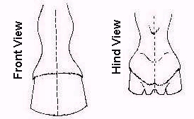
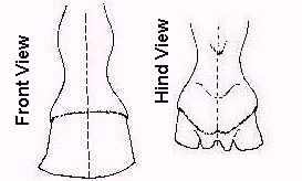
When a horse applies weight on the foot,
changes occur in the shape of the hoof. These
physiological movements are essential to the health of
the foot and the comfort of the animal.

The movements shown in the above
illustrations actually occur almost simultaniously as
weight is placed on the foot. Lateral expansion of
the heel is caused by compression of the plantar
cushion and frog between the foot bones and the
ground. As these elastic structures expand laterally,
they carry with them the lateral cartilages and the
rear portion of the hoof wall. When the foot is
lifted, all of these structures snap back to their
original resting position. Shoeing interferes with
these movements, particularly if the shoe is too big
and the posterior nail is too far backward toward the
heel.
